Skin health education
ABCDE Melanoma Rule: How to Check Your Moles for Skin Cancer
Learn the ABCDE melanoma rule for early skin cancer detection. Understand asymmetry, border, color, diameter, and evolution of moles.
Prepared by NextPath Labs Medical Team
The ABCDE rule is a simple guide to help identify potential melanoma warning signs. This method helps you remember key characteristics to look for when examining moles or skin lesions that might indicate melanoma, the most serious form of skin cancer.
Regular self-examinations of your skin can help detect potential skin cancers in their early stages. When performing these checks, use the ABCDE rule as your guide to identify suspicious moles that should be evaluated by a dermatologist.
Download the original ABCDE guide documentThe ABCDE Rule Explained
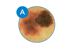
A: Asymmetry
One half of the mole doesn't match the other half. Healthy moles are typically symmetrical. If you were to draw a line through the middle, both sides would match in terms of shape, color, and general appearance. Melanomas often have an irregular shape where the two halves look different.
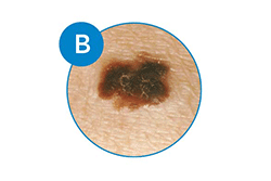
B: Border
The edges are irregular, ragged, notched, or blurred. Normal moles typically have smooth, even borders. Melanomas often have irregular, notched, or scalloped edges that don't form a consistent border. The pigment may appear to spread into the surrounding skin.
C: Color
The color isn't the same throughout the mole. Healthy moles are usually a single shade of brown. Melanomas often display multiple colors within the same lesion, including various shades of brown, black, blue, red, pink, or white. This color variation is a warning sign.
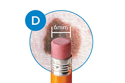
D: Diameter
The mole is larger than 6 millimeters across, about the size of a pencil eraser. Normal moles are typically smaller than 6mm in diameter. Melanomas are often larger than this, though they can be smaller when first detected. Any growth in size should be monitored carefully.
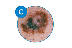
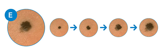
E: Evolving
The mole is changing in size, shape, or color. Healthy moles generally remain stable over time. Melanomas tend to change over time, in size, shape, color, elevation, or symptoms like bleeding, itching, or crusting. Any change in a mole should be professionally evaluated.
When to See a Doctor
If you notice any of the ABCDE signs in a mole, or if you have a mole that's changed in any way, it's important to have it checked by a healthcare professional. While not all suspicious moles are cancerous, early detection of melanoma is crucial for successful treatment.
Remember: Early detection of melanoma is associated with a 5-year survival rate of over 98%. This rate decreases significantly when melanoma is detected at later stages.
Remember, this guide is not a substitute for professional medical advice. Regular skin checks by a dermatologist are recommended, especially for individuals with risk factors such as:
- Fair skin, light hair, or light eyes
- History of sunburns, especially in childhood
- Family or personal history of skin cancer
- Many moles or unusual moles
- Weakened immune system
Recommended Skin Self-Examination Routine
Follow these steps to perform a thorough skin self-examination:
1. Examine your body front and back
Stand in front of a full-length mirror and examine your front, back, and sides with arms raised.
2. Check your arms and hands
Look at the front and back of your arms, underarms, palms, between fingers, and under fingernails.
3. Examine your legs and feet
Check the front and back of your legs, between toes, soles of feet, and around toenails.
4. Check difficult-to-see areas
Use a hand mirror to examine the back of your neck, scalp, back, buttocks, and genital area.
5. Record your findings
Take photos or notes of any moles to track changes over time. DermaVision can help you document and monitor suspicious moles.
Seek immediate medical attention if you notice a mole that is bleeding, oozing, painful, itchy, or has a crusty surface, as these can be signs of more advanced skin cancer.
DermaVision is not a medical device. AI screening is not a diagnosis and cannot rule out skin cancer. It should not replace consultation with a qualified healthcare professional.